What is Dementia?
A common misconception about dementia is that it is itself a disease. In fact, dementia is not a disease, but a rather a group of symptoms. Dementia is a global term that describes chronic and permanent loss of cognitive function severe enough to reduce a person's ability to perform everyday activities.
Here's a quick video to explain more about the causes and symptoms of dementia:
Here's a quick video to explain more about the causes and symptoms of dementia:
Causes
 Feher
Feher
Many conditions and diseases cause dementia. Two of the most common causes of dementia in older people are Alzheimer’s disease and vascular dementia, which is caused by a series of strokes or changes in the brain’s blood supply.
Other conditions that may cause memory loss or dementia include:
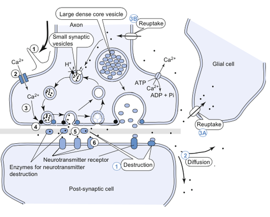
In general, dementia is caused by brain cell damage. The set of brain cells consists of neurons and the neuroglia. functional units of the nervous system are neurons. These cells receive inputs, make decision based on the inputs , and transmit information to other cells. They are characterized by the ability to produce action potentials which allow one part of the cell to communicate with its extreme edges within few milliseconds. Most neurons have thousands of synapses which are connections between neurons. There are two main types of synapses: chemical synapse and electrical synapse. Signals are transmitted across a synapse from one neuron to another neuron neurotransmitters. The general mechanism of neurotransmitters is shown in the picture to the right.
Damages to brain cell interfere with this mechanism and affect the communication between these cells, which in turn will affect the behavior of the brain.
Other conditions that may cause memory loss or dementia include:
- medication side effects
- chronic alcoholism
- tumors or infections in the brain
- blood clots in the brain
- vitamin B12 deficiency
- some thyroid, kidney, or liver disorders
In general, dementia is caused by brain cell damage. The set of brain cells consists of neurons and the neuroglia. functional units of the nervous system are neurons. These cells receive inputs, make decision based on the inputs , and transmit information to other cells. They are characterized by the ability to produce action potentials which allow one part of the cell to communicate with its extreme edges within few milliseconds. Most neurons have thousands of synapses which are connections between neurons. There are two main types of synapses: chemical synapse and electrical synapse. Signals are transmitted across a synapse from one neuron to another neuron neurotransmitters. The general mechanism of neurotransmitters is shown in the picture to the right.
Damages to brain cell interfere with this mechanism and affect the communication between these cells, which in turn will affect the behavior of the brain.
Symptoms
The brain has many distinct regions, each of which is responsible for different functions. Different types of dementia are associated with particular types of brain cell damage in particular regions of the brain. Therefore, symptoms of dementia can vary greatly. Many dementia symptoms start out slowly and gradually get worse. In order to be as dementia, at least two of the following core mental functions must be significantly impaired:
- Memory
- Communication and language
- Ability to focus and pay attention
- Reasoning and judgment
- Visual perception
Mathematical Models
Vascular Dementia
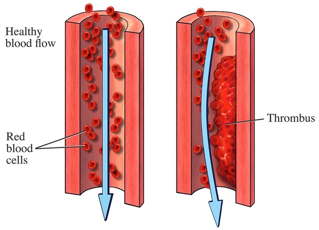
To show the effects of a thrombus (blood clot) in an artery, we take the flow of a healthy internal carotid artery and compare it to the theoretical blood flow of the internal carotid artery with a thrombus.
Some assumptions we will have to make for the theoretical blood flow would be:
- The thrombus causes radius of the artery to decrease, causing the cross section to stay as a circle
- Flow is steady
- flow is laminar
We will be using the formula below to calculate the flow through the artery:
- Q = flow
- ΔP = pressure difference
- R = resistance
Q = ΔP/R (1)
We will be using the formula below to calculate resistance:
- R = resistance
- L = length of the vessel
- η = blood viscosity
- r = inner radius of the vessel
R = 8 L η/π r^4 (2)
|
When we look at a diseased artery and compare it to a healthy one, it is clear that the radius decreases. When looking at equation (2), a decrease in radius causes the resistance to increase. Plugging this new greater resistance back into (1), we find that the flow decreases.
In conclusion, this shows how a thrombus that decreases the radius of an artery will also decrease the flow of the blood. |
http://healthyourpriority.blogspot.com/2013/05/blood-clots-in-lungs.html
|
Alzheimer's Disease
Studies have shown that abnormal deposition of amyloid-β (Aβ) peptides and formation of senile plaques are associated with Alzheimer’s disease (details on the deposition of Aβ plaques will be discussed on pathophysiology section.) Recent studies also suggest that inflammatory activation of microglia, resident innate immune macrophages within brain tissues, may play an important role during the initiation and progression of the disease.
|
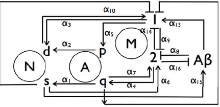
The picture on the right shows the AD mechanism that incorporates feedback influences from surviving and dead neurons (Ns and Nd), quiescent and proliferating astroglia (Aq and Ap), reactive and normal microglia (M1 and M2), and Aβ. (Puri IK 2010)
|
Puri IK 2010
|
The seven rate equations for the cell populations and the number of Aβ molecules in an arbitrary local volume can be expressed:
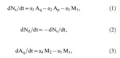
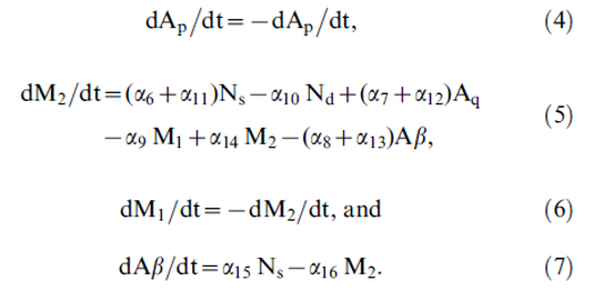
|
Graph (a) represents the population of surviving neurons Ns (black), reactive microglia M1 (red) and Aβ (brown) over 20 years. This graph illustrates equations (6) and (7). Graph (b) shows the population of dead neurons Nd (black) and proliferating astroglia Ap (blue) populations over 20 years.The removal rate ar stabilizes the net number of Ab molecules after three years so that there is only a gradual increase in Nd and corresponding decline in Ns thereafter. The microglia populations are also consequently relatively stable. |

Dynamic simulation of various cell populations
during the progression of Alzheimer’s disease. (Puri IK 2010)
|
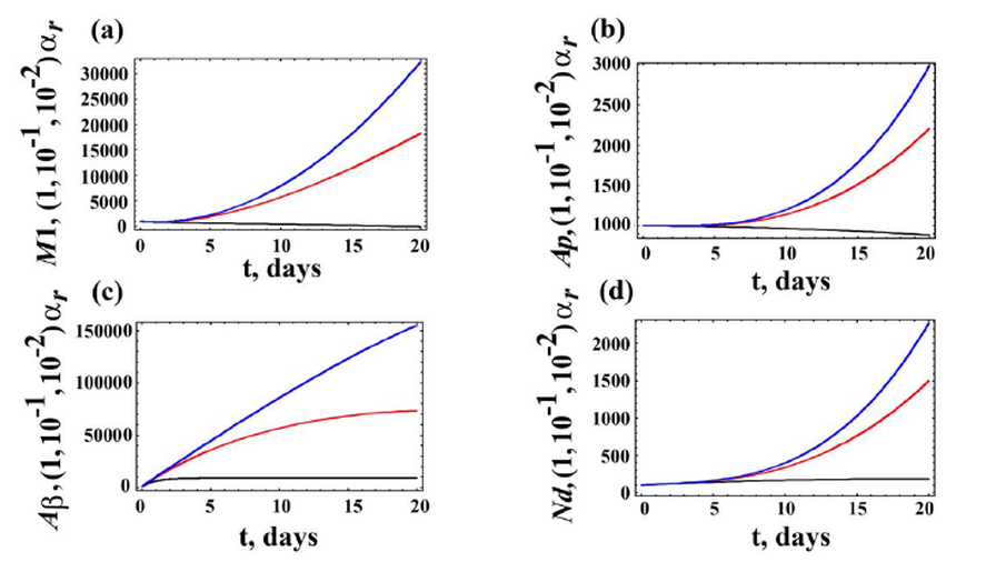
Dynamic variations of cell populations given distinct Ab removal rate. (Puri IK 2010)
The graphs represent the dynamic variations in the (a) M1, (b) Ap, (c) Ab, and (d) Nd populations over 20 years for three values of ar = 16(black), 10216(red), and 10226(blue) the for the Ab removal rate. As ar decreases, there is an increase in europathogenesis so that all four populations increase.
|
The graphs on the right illustrate the dynamic variations in the (a) M1, (b) Nd and (c) Ab populations over 20 years for three values of a13 = 16(black), 106(red), and 506(blue).As α13
increases, M1 and Nd also increase and, consequently, there is an
associated decrease in neuronal survival. This is also illustrated through Eqs.
(1) and (2) of the mathematical model
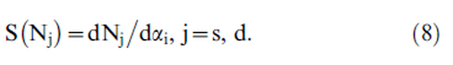
The sensitivities of these cells can be determined by the definition of sensitivity coefficient:
|
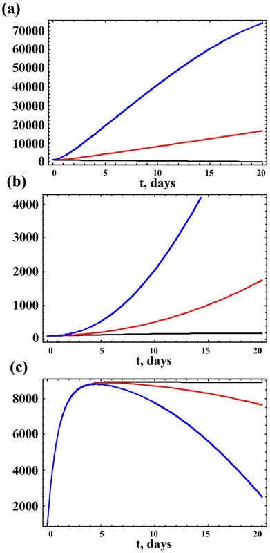
Dynamic variations of cell populations given distinct
impacts of Ab on M1 macrophages (the value of a13).
|
